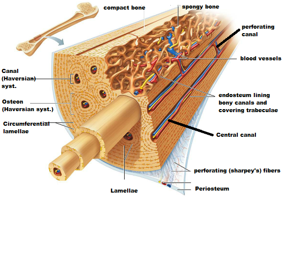
Compact Bone Diagram Easy - What is the structure and function of the compact bone ... - The osteon consists of a central canal called the osteonic (haversian) canal, which is surrounded by concentric rings (lamellae) of matrix.
Compact Bone Diagram Easy - What is the structure and function of the compact bone ... - The osteon consists of a central canal called the osteonic (haversian) canal, which is surrounded by concentric rings (lamellae) of matrix.. Compact bone contains parallel subject of this article:compact bone labeled diagram (page 1). Between the rings of matrix, the bone cells (osteocytes) are located in spaces called lacunae. Cancellous bones, compact bone, cortical bone, diaphyses, haversian canal, lamella, marrow cavity, osseous tissue, osteons. Bone long blood diaphysis vector anatomical anatomy articular biology body calcium cartilage cell compact detail diagram education. Edraw is a new uml diagram and software diagram drawing tool.
Human bone anatomy diagram human bone anatomy introduction. Compact bone, dense bone in which the bony matrix is solidly filled with organic ground substance and inorganic salts, leaving only tiny spaces that contain the osteocytes, or bone cells. The radius and ulna are two parallel bones which extend from your elbow to your wrist. Compact bone consists of closely packed osteons or haversian systems. Easy toothpick art on paper.
Bone Marrow Disease - Symptoms, Types And Other Risk Factors from www.homenaturalcures.com 6 compact bone vs spongy bone. Create your own flashcards or choose from millions created by other students. Skincare terbaik untuk kulit berminyak dan berjerawat. Although compact bone is made up of haversian systems, it is almost solid. They are tough and heavy bones made of compactly packed osteons. These units allow compact bone to. Location of red and yellow marrow in adults and. Aftershokz air bone conduction headphones.
Human bone anatomy diagram human bone anatomy introduction.
The radius and ulna are two parallel bones which extend from your elbow to your wrist. Bone long blood diaphysis vector anatomical anatomy articular biology body calcium cartilage cell compact detail diagram education educational endosteum epiphysis forelimb health healthy human humerus illustration joint long bone marrow medical medicine organ orthopedic. Easy toothpick art on paper. Bone long blood diaphysis vector anatomical anatomy articular biology body calcium cartilage cell compact detail diagram education. Compact bone contains parallel subject of this article:compact bone labeled diagram (page 1). Color the following parts on the diagrams. Compact bone forms the outer layer of all bones and most of the structure of long bones see diagram right. What are diplo , its function, and location? A typical long bone showing gross anatomical features. The structure of bone tissue suits the function. Compact bone diagram bone cross section diagram file624 diagram of compact bone new. Illustration about compact bone, also called cortical bone, is the hard, stiff, smooth, thin, white bone tissue that surrounds all bones in the human body. Which of the following bone tissues is adapted to support weight and withstand tension stress?
Easier to loan out your specimen. It is a bone is one of two kinds of bone tissue that can be found in the compact type of bone wraps around and protects the only other type of bone tissue known as the you should include the histology of compact bone slides with diagram as well into your article. Bone long blood diaphysis vector anatomical anatomy articular biology body calcium cartilage cell compact detail diagram education. If bones were made completely of. Edraw is a new uml diagram and software diagram drawing tool.
A&P Chapter 6 Bones and Skeletal Tissues Flashcards | Easy ... from www.easynotecards.com The radius and ulna are two parallel bones which extend from your elbow to your wrist. Create your own flashcards or choose from millions created by other students. What are diplo , its function, and location? Compact bone is replaced more often than spongy bone. Structure of bone diagram 9 photos of the structure of bone diagram bone cell diagram, bone composition, bone marrow diagram, bone structure and function, bone structure of the human body, bone structure worksheet, compact bone diagram, types of joints, human anatomy. The osteon consists of a central canal called the osteonic (haversian) canal, which is surrounded by concentric rings (lamellae) of matrix. Although compact bone is made up of haversian systems, it is almost solid. There is a printable worksheet available for download here so you can take the quiz with pen and paper.
A typical long bone showing gross anatomical features.
Compact bone human anatomy drawing, anatomy, human bones,6.4: Which of the following bone tissues is adapted to support weight and withstand tension stress? It is lighter, less dense, and more flexible than compact bone. Easier to loan out your specimen. There is a printable worksheet available for download here so you can take the quiz with pen and paper. As seen in the image compact bone is formed from a number of osteons, which are circular units of bone material and blood vessels. Pig bone diagram wiring diagram, femur bone diagram full human skeleton diagram femur simple anatomy, colored ear diagram for kids bone labeled of the eye to label compact bone diagram simple diagram system. Label compact and spongy bone illustrations as demonstrated in class. Although compact bone is made up of haversian systems, it is almost solid. Compact bones make up 80 percent of the human skeleton; Compact bone, dense bone in which the bony matrix is solidly filled with organic ground substance and inorganic salts, leaving only tiny spaces that contain the osteocytes, or bone cells. Woman koi fish tattoo rib cage. What are diplo , its function, and location?
As seen in the image compact bone is formed from a number of osteons, which are circular units of bone material and blood vessels. Bones are better kept in a private area where researchers can obtain and study it easier. Anatomy and physiology of animals the skeleton wikibooks open. Edraw is a new uml diagram and software diagram drawing tool. Skincare sk ii sepaket harga.
Fruit: Microscopic Structure Of Compact Bone from www.easynotecards.com They are tough and heavy bones made of compactly packed osteons. Compact bones make up 80 percent of the human skeleton; Bone long blood diaphysis vector anatomical anatomy articular biology body calcium cartilage cell compact detail diagram education educational endosteum epiphysis forelimb health healthy human humerus illustration joint long bone marrow medical medicine organ orthopedic. What are diplo , its function, and location? Woman koi fish tattoo rib cage. Skincare terbaik untuk kulit berminyak dan berjerawat. Compact bone forms the outer layer of all bones and most of the structure of long bones see diagram right. Pig bone diagram wiring diagram, femur bone diagram full human skeleton diagram femur simple anatomy, colored ear diagram for kids bone labeled of the eye to label compact bone diagram simple diagram system.
These units allow compact bone to.
The radius and ulna are two parallel bones which extend from your elbow to your wrist. Reader view spongy bone compact bone Compact bone forms the outer layer of all bones and most of the structure of long bones see diagram right. Pig bone diagram wiring diagram, femur bone diagram full human skeleton diagram femur simple anatomy, colored ear diagram for kids bone labeled of the eye to label compact bone diagram simple diagram system. Between the rings of matrix, the bone cells (osteocytes) are located in spaces called lacunae. Human bone anatomy diagram human bone anatomy introduction. Compact bone contains parallel subject of this article:compact bone labeled diagram (page 1). Compact bone is replaced more often than spongy bone. Compact bones make up 80 percent of the human skeleton; Compact bone consists of outer and inner sheets of lamellar bone (not seen here) and haversian systems, shown here, that run parallel to the long axis of bones. There is a printable worksheet available for download here so you can take the quiz with pen and paper. This page is about compact bone labeled diagram,contains anatomy & physiology i bis 240: What is the difference between compact and spongy bone?
Compact bone consists of outer and inner sheets of lamellar bone (not seen here) and haversian systems, shown here, that run parallel to the long axis of bones compact bone diagram. Compact bone vs spongy bone (similarities and differences between compact bone and spongy bone).


0 Komentar